This PDF presents Locomotion and Movement through detailed NCERT-style zoology maps. It explains how living organisms move, the difference between movement and locomotion, and the structural and functional systems that make movement possible in humans. The content is highly visual and concept-based, covering muscles, skeletal system, joints, mechanism of muscle contraction, and common movement-related disorders in a structured, exam-focused manner.
I am writing about this topic because Locomotion and Movement is one of those chapters where students often struggle to connect diagrams with theory. These zoology maps bring clarity by linking concepts, labels, tables, and processes on a single platform. For Class 11 students and competitive exam aspirants, understanding this chapter properly is important as it combines biology, anatomy, and physiology in a way that directly reflects NCERT-based questions.
Movement and Locomotion: Basic Concepts
The PDF begins by clearly distinguishing between movement and locomotion. Movement is described as a general characteristic of all living beings, while locomotion refers to voluntary movements that result in a change of place or location. Examples such as walking, running, flying, and swimming are used to show locomotion, while internal movements like blood circulation are classified only as movement.
It also explains why organisms perform locomotion, such as searching for food, escaping predators, finding mates, and reaching breeding grounds, setting a biological context for the chapter.
Types of Movement in Living Organisms
Different types of movement are explained with structure and examples.
Amoeboid movement involves pseudopodia and is seen in Amoeba, leucocytes, and macrophages.
Ciliary movement occurs due to cilia and helps in functions like removing dust from the trachea and movement of ova in the female reproductive tract.
Flagellar movement is seen in organisms like Euglena and spermatozoa.
Muscular movement is the most advanced type and includes movements of limbs, jaws, tongue, and body posture, making it essential for human locomotion.
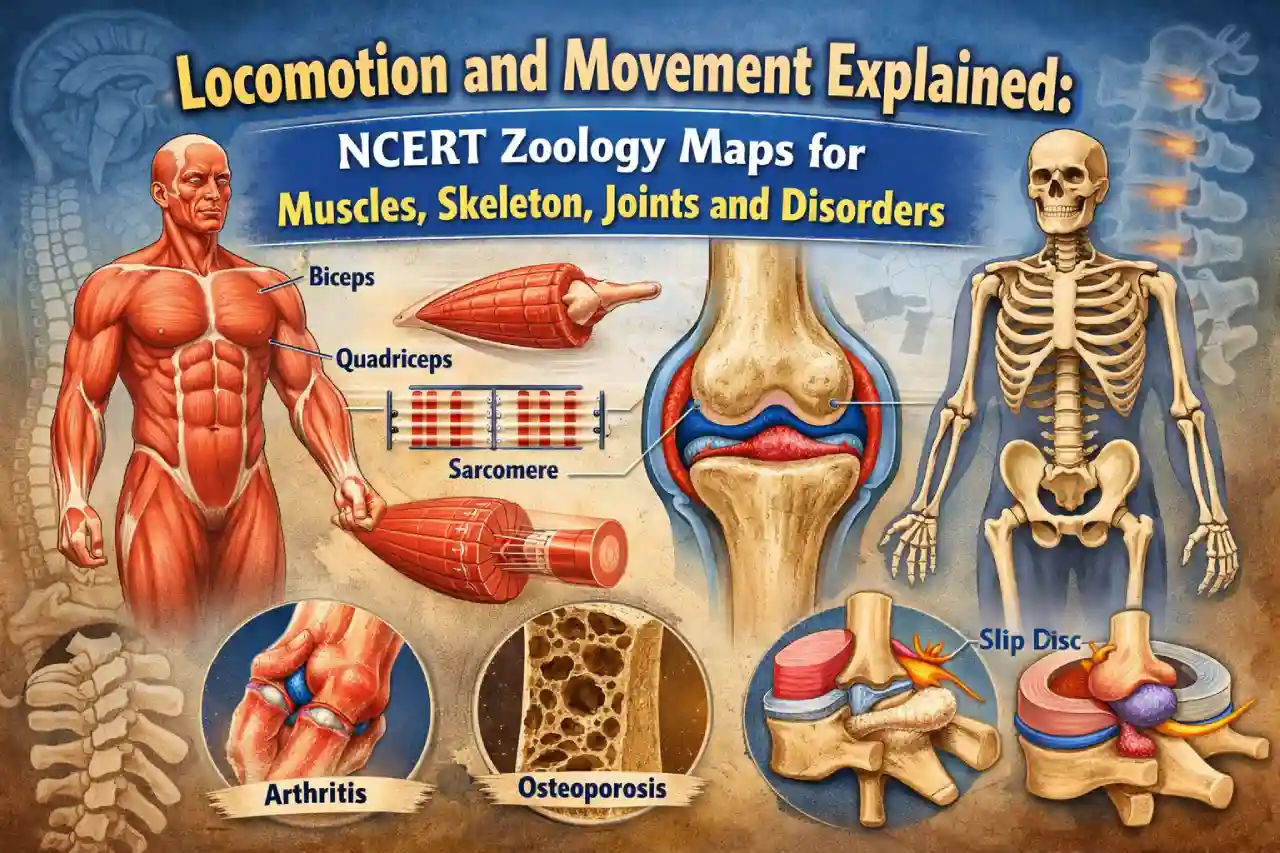
Muscles: Structure, Properties and Types
The PDF explains that muscle tissue is mesodermal in origin and contributes about 40–50 percent of body weight in humans. Key properties of muscles such as excitability, contractility, extensibility, and elasticity are clearly listed.
Muscles are classified into skeletal, visceral, and cardiac muscles based on location, appearance, and regulation. Skeletal muscles are voluntary and striated, visceral muscles are involuntary and smooth, and cardiac muscles are involuntary but striated, forming the muscular tissue of the heart.
Download this Zoology – Map – Samples PDF File: Click Here
Skeletal Muscle Fibres and Myofilaments
A detailed section focuses on skeletal muscle fibres, explaining sarcolemma, sarcoplasm, sarcoplasmic reticulum, and myofibrils. The PDF clearly identifies muscle fibre as the anatomical unit of muscle.
The structure of myofilaments is explained using actin and myosin filaments. Thin filaments consist of actin, tropomyosin, and troponin, while thick filaments are made of myosin. The arrangement of A-band, I-band, H-zone, Z-line, and M-line is explained with clear diagrams, making sarcomere structure easy to understand.
Mechanism of Muscle Contraction
The sliding filament theory is explained step by step. According to the PDF, muscle contraction occurs when thin actin filaments slide over thick myosin filaments. The role of calcium ions, troponin, tropomyosin, ATP, and cross-bridge formation is clearly described.
It explains how acetylcholine at the neuromuscular junction initiates the process, leading to shortening of the sarcomere. Changes in I-band and H-zone with no change in A-band length are highlighted as key exam points.
Skeletal System: Axial and Appendicular Skeleton
The skeletal system is explained as the framework of 206 bones and cartilages. The axial skeleton includes skull, vertebral column, ribs, and sternum, while the appendicular skeleton consists of limb bones and girdles.
The PDF details skull bones, vertebral regions, rib types, and the structure of pectoral and pelvic girdles. Important features like dicondylic skull, true and false ribs, and the longest bone of the body, femur, are clearly mentioned.
Joints and Their Role in Movement
Joints are described as points of contact between bones that enable movement. The PDF classifies joints into fibrous, cartilaginous, and synovial joints based on structure and movement.
Examples such as sutures of the skull, intervertebral joints, knee joint, shoulder joint, and hip joint are used to explain how joints act as fulcrums for movement during muscle action.
Disorders of Muscular and Skeletal System
The PDF concludes with important disorders related to movement. These include myasthenia gravis, muscular dystrophy, tetany, arthritis, gout, and osteoporosis. Causes such as autoimmunity, genetic defects, calcium imbalance, uric acid accumulation, and hormonal changes are clearly linked with their effects on muscles and bones.