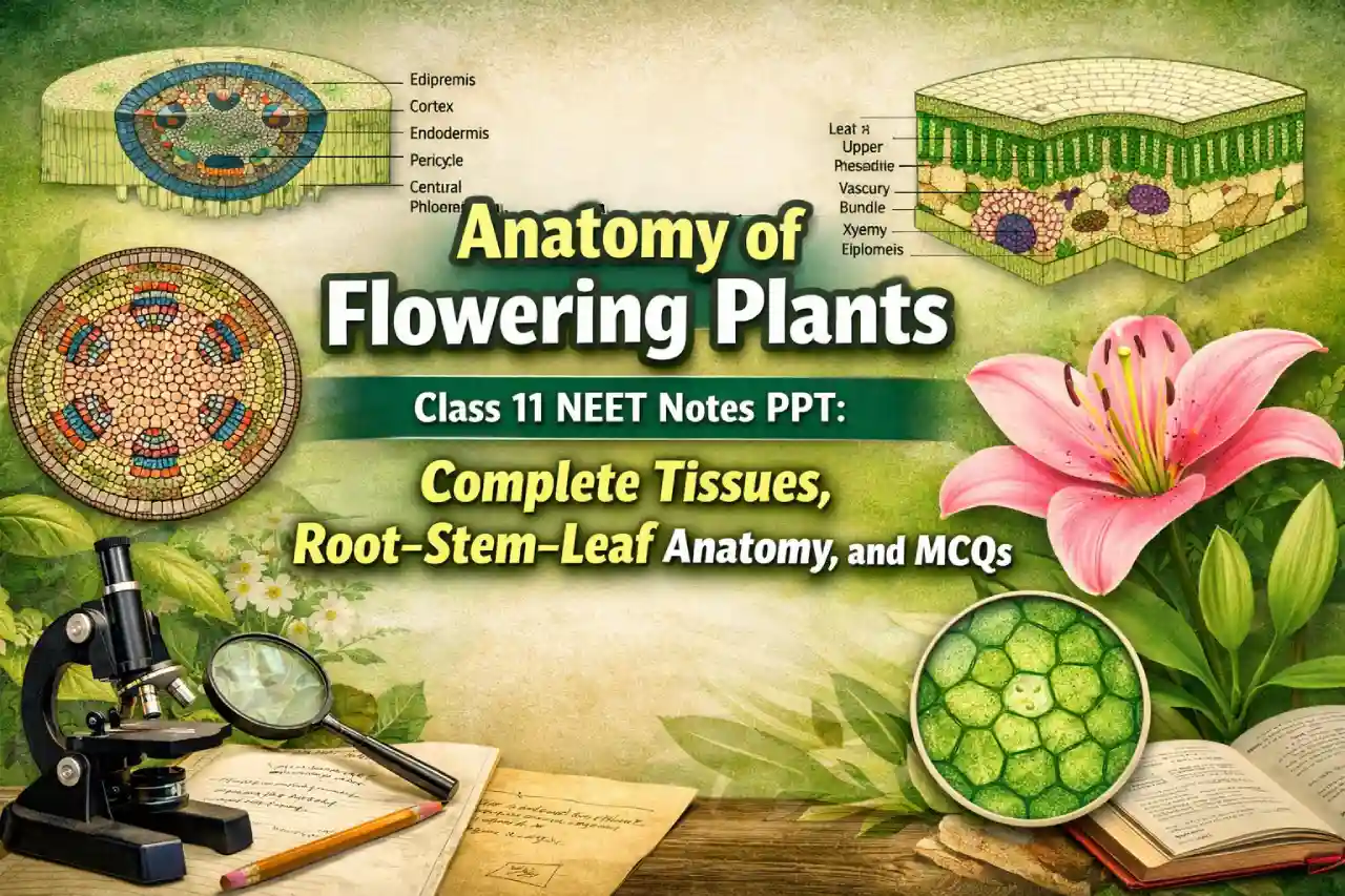
Anatomy of Flowering Plants is a core chapter in Class 11 Biology that explains the internal structure and functional organisation of plants. The uploaded PPT titled Anatomy of Flowering Plant – Class 11 NEET is a well-structured classroom presentation that covers introduction to plant anatomy, tissues, types of tissues, meristematic and permanent tissues, tissue systems, and detailed anatomy of root, stem, and leaf. It also includes a large number of NEET and NCERT-based multiple-choice questions with answers, making it both a concept-building and exam-oriented resource.
I am writing about this PPT because this chapter carries good weightage in NEET and board exams, yet many students struggle to remember structural details and tissue functions. This presentation simplifies complex anatomical features into clear points and diagrams. Knowing what this PPT contains and how to use it can help students revise systematically and score better in Biology.
Introduction to Anatomy and Plant Tissues
The PPT begins by defining anatomy as the study of internal structure and functional organisation of plants. It also mentions that Nehemiah Grew is known as the father of plant anatomy, while K. A. Chaudhary is regarded as the father of Indian plant anatomy.
A tissue is defined as a group of cells having a common origin and performing a common function. Based on their capacity to divide, tissues are classified into:
- Meristematic tissue
- Permanent tissue
This basic classification sets the foundation for the entire chapter.
Meristematic Tissue
Meristematic tissues are composed of actively dividing, undifferentiated cells responsible for plant growth. The PPT explains that the term “meristem” is derived from the Greek word meristos, meaning divided.
Meristems are classified based on origin and position:
- Primary meristem
- Secondary meristem
- Apical meristem
- Intercalary meristem
- Lateral meristem
Apical meristems are present at root and shoot tips and help in primary growth. Intercalary meristems occur at internodes or leaf bases and are responsible for elongation, especially in grasses. Lateral meristems such as vascular cambium and cork cambium are responsible for secondary growth.
Important features of meristematic cells mentioned in the PPT include:
- Large nucleus
- Dense cytoplasm
- Thin primary cell wall
- Absence of vacuoles and intercellular spaces
Permanent Tissues
Permanent tissues are formed from meristems and have lost the capacity to divide. They are classified into:
- Simple permanent tissues
- Complex permanent tissues
Simple Permanent Tissues
Parenchyma, collenchyma, and sclerenchyma are described in detail.
Parenchyma forms the major component of plant organs. Cells are isodiametric with thin cellulosic walls. Functions include photosynthesis, storage, and secretion.
Collenchyma is present below the epidermis in dicot stems and petioles. Cells have unevenly thickened corners due to cellulose, hemicellulose, and pectin deposition. It provides mechanical support and sometimes helps in photosynthesis.
Sclerenchyma cells are dead with thick, lignified walls. They provide strength. Two types are explained:
- Fibres (elongated and pointed)
- Sclereids (stone cells found in nut shells, guava pulp, pear, sapota, and seed coats)
Complex Permanent Tissues
Complex tissues consist of more than one type of cell and are mainly conducting tissues.
Xylem elements include:
- Tracheids
- Vessels
- Xylem fibres
- Xylem parenchyma
Tracheids and vessels are main water-conducting elements. Vessels are characteristic of angiosperms and absent in gymnosperms.
Phloem elements include:
- Sieve tubes
- Companion cells
- Phloem parenchyma
- Phloem fibres
Sieve tubes have perforated end walls called sieve plates and lack nucleus at maturity. Companion cells control their functions. Phloem fibres are sclerenchymatous and used commercially (jute, flax, hemp).
Download this PPT File: Click Here
Tissue Systems
The PPT explains three tissue systems:
- Epidermal tissue system
- Ground tissue system
- Vascular tissue system
These systems together form the structural framework of plant organs.
Anatomy of Root
Dicot Root
The internal structure includes epiblema, cortex, endodermis with Casparian strips, pericycle, vascular bundles, and pith.
Monocot Root
Structure is similar to dicot root, but xylem bundles are more than six (polyarch) and pith is large and well developed. Monocot roots do not undergo secondary growth.
Anatomy of Stem
Dicot Stem
Epidermis, cortex (with hypodermis, cortical layers, and endodermis), pericycle, vascular bundles arranged in a ring, medullary rays, and pith are present.
Monocot Stem
Has sclerenchymatous hypodermis, scattered vascular bundles with bundle sheath, large parenchymatous ground tissue, conjoint and closed vascular bundles, and absence of phloem parenchyma.
Anatomy of Leaf
Dicot Leaf (Dorsiventral)
Shows epidermis, mesophyll (palisade and spongy parenchyma), and vascular bundles.
Monocot Leaf (Isobilateral)
Stomata present on both surfaces, mesophyll not differentiated, presence of bulliform cells, and parallel venation.
NEET and NCERT-Based MCQs
Throughout the PPT, multiple-choice questions are provided after each topic with correct answers. These questions help students test conceptual clarity and understand how questions are framed in NEET.
How Students Can Use This PPT
- Study theory topic-wise
- Revise diagrams and structures
- Practise MCQs regularly
- Use it for quick revision before exams